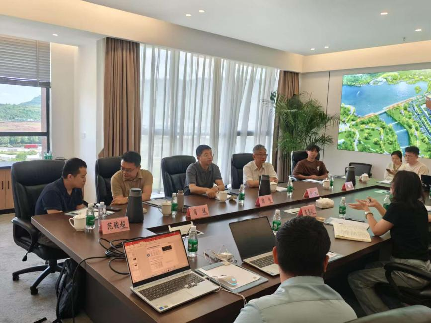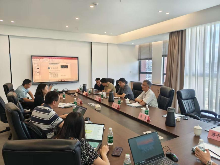
Lecture Review | Fang Bin: "Research on new technology of semantic segmentation of medical images" and "Recognition of surgical video process based on deep learning"; Fan Junchao: "Super-resolution microscopy of living cells"
Lecture review
Lecture 1: Research on new technology of semantic segmentation of medical images
Lecture 2: Deep Learning-based surgical video process recognition

Introduction to the speaker
Fang Bin,Professor and doctoral supervisor of the School of Computer Science and Technology of Chongqing University, and director of the Institute of Pattern Recognition and Information Processing. The main research directions are: artificial intelligence, computer vision, pattern recognition, medical image processing, etc. IEEE Senior Member, a scholar in Bayu, Chongqing, and an outstanding talent in the new century of the Ministry of Education. Deputy editor-in-chief of SCI international journal "IJPRAI" and invited editor of SCI international journal "MPE". Member of the Chongqing CPPCC, Vice Chairman of the Chongqing Digital Medicine Society, and Secretary-General of the Chongqing Cognitive Science Society. "Credited Computing" academic leader of the Ministry of Education's key laboratory and "Software Theory and Technology" academic leader of the Chongqing Key Laboratory. He won 1 first prize in the Ministry of Education Natural Science Award and 2 second prizes in the Chongqing Natural Science Award. It undertakes more than 30 important projects such as the 973 project of the Ministry of Science and Technology, the Support Plan Project of the Ministry of Science and Technology, and the Natural Science Foundation of the Fund of the Committee. More than 200 SCI papers have been published. More than 10 authorized invention patents have been obtained.
Lecture one main content
Semantic segmentation of brain choroid plexus images is an important method of tissue structure recognition. Although it provides key positioning information for neuroimage analysis, due to the complex image and blurred labeling, there are problems of segmentation accuracy and efficiency, which can easily lead to incomplete identification of fine structures or unclear boundaries. This technical defect directly leads to a decrease in data reliability when segmenting the fine structure of the brain choroid plexus in an automated and high-precision manner. Long-term application data shows that the accuracy and robustness of traditional semantic segmentation algorithms in choroid plexus segmentation are far inferior to the ideal state.
The research found that training data based on manual annotation has significant limitations in complex scenarios: fuzzy boundary areas are easily affected by subjective judgment, resulting in small structure missed segmentation and background missed segmentation, and systematic deviations are introduced in algorithm training. High-quality choroidal plexus segmentation requires balancing model complexity, labeling accuracy and generalization capabilities. This points out the direction for algorithm optimization: developing smart algorithms that are more discriminant and able to capture structural priors can effectively reduce segmentation errors and improve the credibility of results.
In order to break through the limitations of traditional semantic segmentation methods, Professor Fang Bin’s team innovatively developed the brain choroid plexus intelligent segmentation algorithm. This breakthrough not only solves the core problems of accuracy and generalization capabilities in choroid plexus segmentation, but also opens up a new paradigm in the field of medical image segmentation. The importance of this research is reflected in multiple dimensions: First, the accuracy and efficiency advantages of segmentation are organically combined through optimization strategies based on multi-scale feature fusion and structural constraints, and the deep collaboration between deep learning architecture and anatomical prior knowledge is realized for the first time, providing an innovative template for the automation and refined recognition of brain choroid plexus; Second, the spatial distribution and morphological characteristics of choroid plexus are used as segmentation strategies with strong prior constraints, breaking through the limitations of traditional algorithms that simply rely on pixel-level annotations, and cascade improvement of segmentation results (boundary fit, small structural integrity, anti-noise interference ability) is achieved from the algorithm level. This innovation not only significantly enhances the clinical practical value of choroid plexus segmentation, but also provides new research ideas and solutions for the intelligent structure analysis of medical images, and has an important impact on promoting neuroscience research and related diseases diagnosis and treatment.
Lecture 2 main content
Intelligent analysis of surgical videos is a key technology for clinical diagnosis and treatment and surgical training, but its accurate analysis in dynamic surgical scenarios is often limited by the decline in video quality caused by tissue fog, blood occlusion and dynamic blur. These interfering factors cause the loss or distortion of key information (such as instrument-tissue interaction, critical anatomy), seriously affecting the robustness and depth of analysis in complex surgical scenarios.
The research found that the remaining effective information in fuzzy, low-contrast videos has unique patterns, but is easily masked by noise and distorted structure, resulting in rapid degradation of detailed features and semantic information. This reveals that the key to improving analytics abilities lies in breaking through the dependence on the quality of original videos. To this end, Professor Fang Bin’s team innovatively developed intelligent analysis technology for surgical video defog removal. The core of this technology lies in using a defog enhancement algorithm based on multi-scale feature fusion and physical model guidance to significantly improve the clarity of surgical videos. By effectively suppressing interference such as fog and blood stains, the key details that were originally covered are clearly presented. At the same time, this technology makes full use of the space-time continuity and anatomical prior knowledge of surgical scenes as constraints, breaking through the limitations of traditional algorithms relying solely on pixel clarity. This breakthrough progress solves the core problem of information loss in intelligent analysis of surgical videos, bringing cascade improvement: First, the images are clearer, effectively removing mist and blood stains, and significantly improving the video quality; Second, the analysis is more accurate, and the clear images greatly improve the integrity and accuracy of the identification of key steps (such as instrument operation and anatomical structure positioning); Third, the algorithm is more robust, and in complex interference environments, the anti-interference and timing consistency of the analysis results are significantly enhanced; Fourth, efficiency and practicality are improved, providing a more reliable foundation for high-precision real-time intelligent analysis of the entire process of the surgery. This technology not only significantly enhances the clinical practical value of intelligent surgical video analysis, but also provides new ideas for surgical navigation and postoperative evaluation, but also opens up a new paradigm in the field of computer-assisted surgery, which has an important impact on promoting the development of precision surgery and intelligent surgery.
Lecture 3: Super-resolution microscopy of living cells

Introduction to the speaker
Fan Junchao,Associate Professor and PhD Supervisor at Chongqing University of Posts and Telecommunications, and a young scholar in Bayu, Chongqing. He is mainly engaged in research on multimodal image perception and characterization, and his achievements are applied to biomedical image analysis and image fusion. He has published more than ten papers as (co-)first author or correspondent author in high-level journals such as Nature Biotechnology and Nature Communications. He presided over 1 key research and development project of the Ministry of Science and Technology, 1 National Natural Science Foundation Youth Project, and 3 provincial and ministerial projects, participated in 1 original exploration project of the National Natural Science Foundation, and was authorized to 2 national invention patents. The Heisen structured light illumination fluorescence microscope developed is currently the fastest and most sensitive live cell super-resolution imaging technology, reaching the world's leading level and winning scientific research awards such as the "Top Ten Progress in China's Optics" in 2018.
The three main contents of the lecture
Fluorescence Light Microscopy is a commonly used optical imaging method in biomedical research and can provide key information. However, when super-resolution imaging of living cells, due to the influence of phototoxicity and photobleaching effects, the imaging time and resolution are limited, resulting in incomplete observation or distortion of dynamic processes of living cells, which has become a key bottleneck in the application of technology. This technical defect causes data quality to decline when long-term and high-resolution observation of fine structures such as organelles dynamics in living cells. Long-term application data show that traditional super-resolution technology has a significant gap with the ideal state in terms of continuous observation ability and information revelation of live cell imaging.
The study found that cells surviving under high doses of light showed a unique biological response. These cells activate the photodamage stress pathway under strong light stimulation, manifested as rapid quenching of fluorescent proteins and a decrease in cell viability, which marks the widespread photochemical damage in the cells. Under normal circumstances, live cell imaging requires balancing light intensity, exposure time and resolution. This discovery provides an important opportunity for imaging technology optimization - by developing imaging algorithms with lower photon requirements and higher sensitivity, it can effectively suppress the photodamage effect and extend the observable window of living cells.
In order to break through the limitations of traditional light illumination technology, Professor Fan Junchao's team innovatively developed Hessian-SIM structured light illumination fluorescence microscope (Hessian-SIM). This breakthrough not only solves the core problems of phototoxicity and speed in super-resolved live cell imaging, but also opens up a new paradigm in the field of biomedical microscopy. The importance of this research is reflected in multiple dimensions: First, the sensitivity and speed advantages of super-resolution imaging are organically combined through the deconvolution algorithm based on Hessian matrix, and the deep collaboration between computational optics and artificial intelligence algorithms is realized for the first time, providing an innovative template for long-term and high-resolution imaging of living cells; Second, the spatial continuity of biological structures is used as a reconstruction strategy for prior knowledge, breaking through the limitations of traditional algorithms' simple dependence on the number of photons, and the cascade improvement of image quality (resolution, signal-to-noise ratio, and anti-artifact capability) is achieved from the algorithm level; Third, the technical path has excellent performance: the super-resolution image with minimal artifacts (88 nm resolution) is obtained without a photon dose of 10% of the traditional structured light illumination micromirror (SIM), and the time resolution is as high as 188. Hz; its high sensitivity supports the use of submillisecond pulse excitation combined with long dark recovery time, significantly reducing photobleaching, achieving time-lapse super-resolution imaging of fine structures (such as actin filaments) in living cells for up to one hour, and can clearly analyze the dynamic structural changes of mitochondrial cristae. This innovation not only significantly improves the living application capabilities of super-resolution microscopy imaging, but also provides new research ideas and solutions for super-resolution microscopy of living cells, which is of great significance to promoting dynamic observation and research in life sciences.
- About Us
-
Research Platform
- Major Disease Sample Database
- Innovative Drug Verification And Transformation Platform
- Experimental Animal Center
- Life And Health Future Laboratory
- Biomedical Imaging Platform
- Cell Multi-Omics Platform
- Pathology Technology Platform
- Bioinformatics Research And Application Center
- Jinfeng Pathology Precision Diagnosis Center
- Research Team
- Information Center
- Join Us


 渝公网安备50009802002274