
Lecture review | intercellular communication and tumors

lecture title
"intercellular communication and tumors"
introduction to the keynote speakers
professor kang tiebang, deputy director and pi of the sun yat-sen university cancer center/national key laboratory of malignant tumor prevention and control in south china, professor, doctoral supervisor, "changjiang scholar" distinguished professor of the ministry of education, winner of the national science fund for outstanding youth, tumor cell of the chinese society of cell biology president of the biology branch. in 2003, he obtained a phd in biochemistry from bielefeld university in germany. from 1998 to 2008, he engaged in cell biology and oncology research in the united states and germany. in may 2008, he returned to china full-time (sun yat-sen university's "hundred talents plan" introduced talents). it has obtained many projects including key projects of the national natural science foundation of china, “973” project projects, and national key research and development plans. he has long been engaged in research on the regulation and intervention of tumor cells and their extracellular vesicles. he has published 50 papers as the corresponding author in mainstream international magazines such as nat cell biol, nat cancer, cell res, sci adv, j clin invest, adv sci, and nat commun. multiple articles.
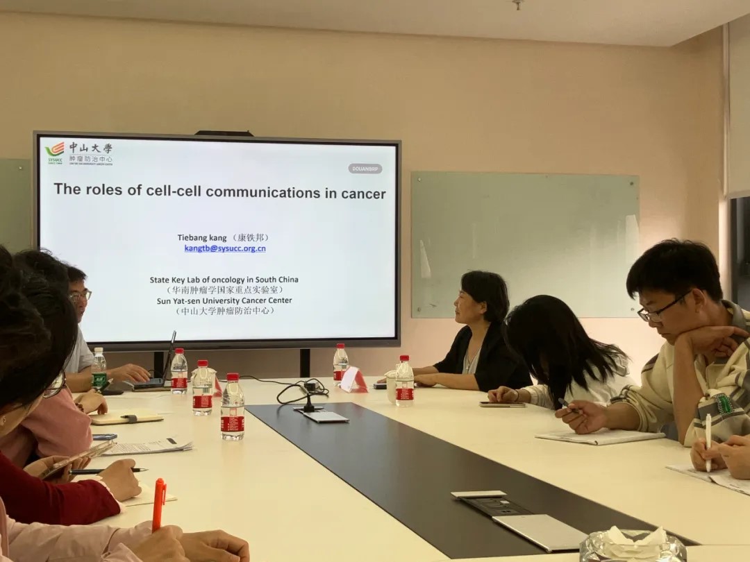
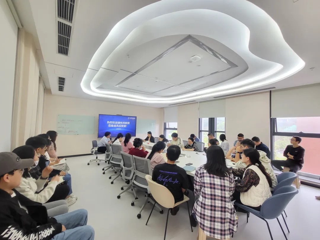
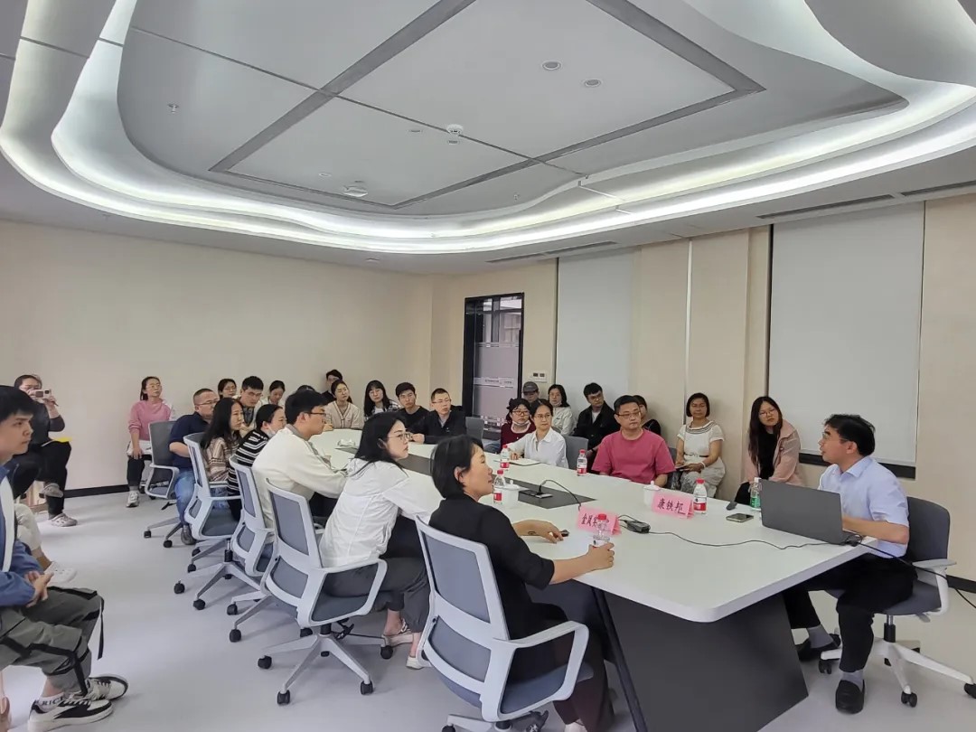
professor kang tiebang was invited to jinfeng laboratory to give academic lectures
key points of the lecture
extravesicles are found in biological fluids and play a role in intercellular communication, allowing cells to exchange proteins, lipids, genetic material, amino acids, and metabolites. external vesicles are a group of heterogeneous membrane structures derived from cells, mainly including exosomes and microvesicles (mvs), which originate from the endosomal system and shedding of the plasma membrane respectively. it has been documented that exosomes and microvesicles contain many cell membrane proteins. during the maturation of endosomes into multivesicular bodies (mves), intraluminal vesicles (ilvs) are formed in the lumen of exosomes, and the multivesicular bodies fuse with the cell surface, and then the intraluminal vesicles are vesicles, secreted outside the cells to become exosomes.
professor kang's team identified an escrt-independent exosome pathway marked and controlled by rab31, revealing the dual functions of rab31 during exosome biogenesis: driving the formation of ilvs and preventing the degradation of mves. a variety of rtks activate rab31 through phosphorylation. activated rab31 drives mves membrane budding to form ilvs by binding to flots proteins in lipid raft microdomains. at the same time, rab31 recruits tbc1d2b to the surface of mves to inactivate rab7 to inhibit the fusion of mves and lysosomes. , thus facilitating the fusion of mves and cell membranes to release ilvs to form exosomes. this study reorganized and defined the molecules related to exosome biogenesis, taking a key step towards better understanding the complex heterogeneity of exosome biogenesis (cell res 2021 feb;31(2):157-77) .
professor kang also shared with the laboratory researchers a detailed explanation of the team's multiple scientific research results in recent years, including a new organelle (rafeesome) that mediates the release of r-ev (rab22a-induced extracellular vesicles). rafeesome can regulate activation intercellular transfer of interferon gene-stimulating factors, while r-ev containing activated interferon gene-stimulating factors can activate and propagate anti-tumor immunity. in summary, this study is helpful to elucidate the process of secretion of the endoplasmic reticulum membrane protein interferon gene stimulating factor out of the cell, and also provides a new perspective for understanding the cellular communication between organelle membrane proteins (cell res. 2022 dec;32(12) ):1086-104).
the team also discovered a new class of exon-intron fusion genes (all known traditional fusion genes are exon-exon), consisting of rab22a (exon 1 and 2, (i.e. 1-38aa) is connected to multiple different introns or non-coding regions, thereby encoding a new fusion protein rab22a-neofs. this type of fusion protein can bind to smggds607, promote the activation of rhoa, and promote the lung development of osteosarcoma. metastasis (nat cell biol, 2020). then, taking the rab22a-neof1 fusion protein as an example, it was found that its k7 acetylation plays a key role in the function of the fusion protein (theranostics, 2020); this fusion protein is secreted into exosomes in a traditional way to remodel tumor microorganisms. immune cells and tumor cells in the environment promote lung metastasis of osteosarcoma (signal transduct target ther, 2021). rab22a-neof1 fusion protein can be ubiquitinated and degraded by autophagy lysosomes. this process can be regulated by phosphorylation. it has been found that the targeted drugs sorafenib and regorafenib being used clinically can induce rab22a. -degradation of the neof1 fusion protein and therefore can be used to treat osteosarcoma patients who are positive for this fusion gene (adv sci (weinh). 2023 feb;10(5):e2205483).
the team recently discovered that under ifnγ stimulation, kat8-irf1 in tumor cells enhances the molecular mechanism of pd-l1 expression by forming condensates with the function of promoting transcription; found that condensate formation can enhance the catalytic rate of kat8's acetylation of irf1; developed blocking peptides that inhibit aggregate formation and demonstrate antitumor activity. this study suggests that tumor-related biological macromolecule aggregates may be a new target for tumor treatment and are worthy of in-depth exploration (nat cancer 2023 mar;4(3):382-400).

 渝公网安备50009802002274
Number
023-68059903
Email:Jinfeng@jflab.ac.cn
Address:
No.313, Bella Road Chongqing High-tech Zone
Postal code: 401329
- About Us
-
Research Platform
- Major Disease Sample Database
- Innovative Drug Verification And Transformation Platform
- Experimental Animal Center
- Life And Health Future Laboratory
- Biomedical Imaging Platform
- Cell Multi-Omics Platform
- Pathology Technology Platform
- Bioinformatics Research And Application Center
- Jinfeng Pathology Precision Diagnosis Center
- Research Team
- Information Center
- Join Us