
Conference review | [microimaging technology frontier exchange conference] cutting-edge technology blessing to help produce scientific research results
microscopic imaging technology has always been an important driving force for the development of the biomedical field. biological microscopic imaging technology has important applications in disease diagnosis, basic research on molecular mechanisms, and drug target discovery. in order to promote the mastery and application of biological microscopic imaging technology, on march 27, jinfeng laboratory and carl zeiss held seminar on cutting-edge microscopy imaging technology.
zhao chengjian, a professor at the state key laboratory of biotherapy at sichuan university, and zhang yan, a senior application engineer at zeiss, gave wonderful academic speeches to the scientific and technical workers present, focusing on the application of microscopic imaging technology and giving a detailed introduction. the exchange meeting attracted more than 70 scientific and technological workers from jinfeng laboratory, bgi, chongqing international institute of immunology, chongqing brain and intelligence science center, the second affiliated hospital of army medical university and other units to exchange, share and explore together. cutting edge.
"application of multiplex immunofluorescence staining (cyclic mif) in assessment of tumor immune microenvironment and precision tumor immunotherapy"
zhao chengjian, professor at the state key laboratory of biotherapy, sichuan university
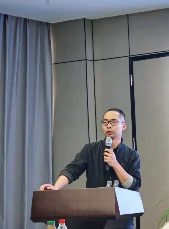
professor zhao chengjian introduced the cyclic mif technology independently developed by the molecular and cellular imaging platform of the national major science and technology infrastructure for translational medicine (sichuan). this technology can realize full-process interoperability from upstream sample preparation, midstream imaging, to downstream data analysis. based on the accurate labeling of numerous immune cell groups in samples, the ai data analysis model is used to display a panoramic map of the tumor microenvironment, revealing information such as cell grouping, interaction, tumor classification, and prognosis, providing a reliable basis for precise treatment. the above-mentioned spatial multi-omics analysis has application potential in precision tumor treatment.
"biological microscopy imaging cutting-edge applications and case sharing - multi-dimensional and cross-modal support for life sciences and medical research dreams"
zhang yan, senior technical expert of zeiss microscope
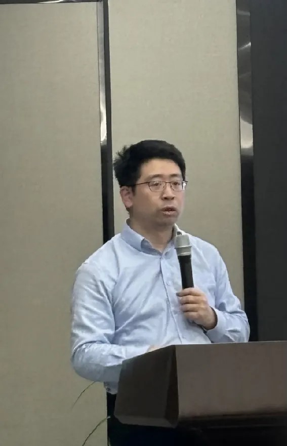
expert zhang yan explained spatial multi-omics research from various aspects such as application examples, technical points and development directions, and expressed the need for comprehensive application of multi-dimensional, cross-modal microscopic imaging and image processing technology.
it requires the accuracy of "seeing", from macroscopic, mesoscopic to microscopic; it requires the three-dimensional "connection" to realize the effective correlation of macro, mesoscopic and microscopic results; it requires the huge "calculation", and analyzes and calculates based on imaging big data , and finally get a highly three-dimensional, integrated and reliable result. zeiss will provide multi-dimensional, cross-modal overall solutions to help jinfeng laboratory's scientific and technological innovation work achieve high-quality development.

the scientific and technological workers participating in the meeting raised a number of technical issues in practice based on their own research directions, and the two experts answered them in detail. this exchange meeting inspired participants' interest in cutting-edge microscopy imaging technology, deepened scientific workers' understanding of high-end microscopy imaging solutions, continuously strengthened the experimental skills of laboratory researchers, and assisted the output of scientific research results.

 渝公网安备50009802002274
Number
023-68059903
Email:Jinfeng@jflab.ac.cn
Address:
No.313, Bella Road Chongqing High-tech Zone
Postal code: 401329
- About Us
-
Research Platform
- Major Disease Sample Database
- Innovative Drug Verification And Transformation Platform
- Experimental Animal Center
- Life And Health Future Laboratory
- Biomedical Imaging Platform
- Cell Multi-Omics Platform
- Pathology Technology Platform
- Bioinformatics Research And Application Center
- Jinfeng Pathology Precision Diagnosis Center
- Research Team
- Information Center
- Join Us